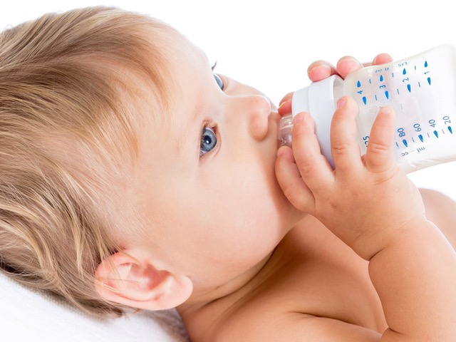
The colonization of the gastrointestinal tract by intestinal bacteria occurs primarily at birth through the mother’s skin and vaginal microbiota (at this point, the microbiota is dominated by Enterobacteriaceae and Staphylococcus).1 The composition of this immature microbiota is temporary because it will evolve significantly during the first months of life, first as a result of lactation (microbiota now dominated by Bifidobacterium)2 and then weaning (primarily Firmicutes and Bacteroidetes),3,4 until finally a relatively stable microbiota is achieved (in the absence of disease) around the age of three, when solid and varied food plays a significant role in stabilizing the microbiota.5
There are a number of factors that modify the composition of the stable intestinal microbiota (type of delivery, breastfeeding versus formula, use of antibiotics, timing of the introduction of solid food, environment, hygiene, etc.).2,6–9 These changes (or modifications) may have an effect on the development of the immune system (changes to immune cells and their messengers, the cytokines),10 thus leading to immune-related diseases such as, for example, food allergies, dermatitis (or eczema) and asthma.11–13 Recent studies have shown dysbiosis (an imbalance in the composition of the microbiota) in these diseases. A decrease in Bacteroides and an increase in Anaerobacters have been identified in food allergies,14 a decrease in Bacteroidetes and Bifidobacterium and an increase in Enterobactiaceae is associated with dermatitis15 and a decrease in the abundance of Bifidobacterium, Akkermensia and Faecalibaterium with asthma.16 In recent years, the excessive hygiene hypothesis (reduced exposure to germs and parasites) has emerged. Numerous studies have shown the rather negative impact of an aseptic environment on the development of allergies. The most at-risk populations (associated with reduced exposure to bacteria) are those that live in an excessively “disinfected” environment. In keeping with these results, certain studies have been able to identify an isolated bacteria (Acinetobacter lwoffii F78) in rural agricultural environments that has a protective effect on allergic mechanisms through its action on immune cells.17
Our current understanding of the bacterial mechanisms underlying these diseases remains incomplete and further analyses will be needed to deepen our understanding of the involvement of the gut microbiota in these pathophysiologies.
Bibliography
- Matsuki, T. et al.A key genetic factor for fucosyllactose utilization affects infant gut microbiota development. Nat. Commun.7, 11939 (2016).
- Mitsuoka, T. Development of functional foods. Biosci. Microbiota Food Health33, 117–128 (2014).
- Vallès, Y. et al.Microbial succession in the gut: directional trends of taxonomic and functional change in a birth cohort of Spanish infants. PLoS Genet.10, e1004406 (2014).
- Palmer, C., Bik, E. M., DiGiulio, D. B., Relman, D. A. & Brown, P. O. Development of the human infant intestinal microbiota. PLoS Biol.5, e177 (2007).
- Yatsunenko, T. et al.Human gut microbiome viewed across age and geography. Nature486, 222–227 (2012).
- Dominguez-Bello, M. G. et al.Delivery mode shapes the acquisition and structure of the initial microbiota across multiple body habitats in newborns. Proc. Natl. Acad. Sci. U. S. A.107, 11971–11975 (2010).
- Azad, M. B. et al.Gut microbiota of healthy Canadian infants: profiles by mode of delivery and infant diet at 4 months. CMAJ Can. Med. Assoc. J. J. Assoc. Medicale Can.185, 385–394 (2013).
- Tanaka, S. et al.Influence of antibiotic exposure in the early postnatal period on the development of intestinal microbiota. FEMS Immunol. Med. Microbiol.56, 80–87 (2009).
- Fouhy, F. et al.High-throughput sequencing reveals the incomplete, short-term recovery of infant gut microbiota following parenteral antibiotic treatment with ampicillin and gentamicin. Antimicrob. Agents Chemother.56, 5811–5820 (2012).
- Francino, M. P. Early development of the gut microbiota and immune health. Pathog. Basel Switz.3, 769–790 (2014).
- Lynch, S. V. & Boushey, H. A. The microbiome and development of allergic disease. Curr. Opin. Allergy Clin. Immunol.16, 165–171 (2016).
- Blázquez, A. B. & Berin, M. C. Microbiome and food allergy. Transl. Res. J. Lab. Clin. Med.179, 199–203 (2017).
- Inoue, Y. & Shimojo, N. Microbiome/microbiota and allergies. Semin. Immunopathol.37, 57–64 (2015).
- Ling, Z. et al.Altered fecal microbiota composition associated with food allergy in infants. Appl. Environ. Microbiol.80, 2546–2554 (2014).
- Abrahamsson, T. R. et al.Low diversity of the gut microbiota in infants with atopic eczema. J. Allergy Clin. Immunol.129, 434–440, 440.e1–2 (2012).
- Fujimura, K. E. et al.Neonatal gut microbiota associates with childhood multisensitized atopy and T cell differentiation. Nat. Med.22, 1187–1191 (2016).
- Debarry, J., Hanuszkiewicz, A., Stein, K., Holst, O. & Heine, H. The allergy-protective properties of Acinetobacter lwoffii F78 are imparted by its lipopolysaccharide. Allergy65, 690–697 (2010).


